Most melanomas are darkly pigmented, but amelanotic (pink) melanomas are possible and are. More rarely, melanomas are found in other parts of the eye.

Anterior Uveal Melanocytic Neoplasia Clinician's Brief
Eyelid tumors in dogs are usually benign, such as sebaceous adenomas and sebaceous epitheliomas.
Uveal melanoma in dog eyelid. Uveal melanomas usually arise from anterior uveal tract (iris or ciliary body). This type of tumor can be both benign or malignant; Female dogs, german shepherds, and labrador retrievers are documented to be predisposed
A mongrel male dog of three years old was referred to the seoul national university veterinary teaching hospital following a one month history of glaucoma. Additionally, as the mass grows, fills the eye and variably distorts or destroys the intraocular structures, differentiation between an originally flat, irial mass or raised, solitary mass is clinically. One would think that our furry friends would be protected from the harmful effects of the sun;
Uveal melanocytic tumors may be benign (melanocytoma) or. Most uveal tumors arise from the iris or ciliary body (part of the wall of the eye that makes the fluid that fills the eye). Ultrasonography and computed tomography revealed a mass with.
These melanocytic tumors are a neoplastic proliferation of the uveal melanocytes and involve the iris, ciliary body, and, rarely, the choroid (2,4,5). Canine uveal cysts are usually benign, but they can also be an initial sign of a progressive blinding disease know as pigmentary uveitis. The most typical site for melanoma tumors to take place in the dog remains in the mouth.
Uveal melanomas are the most common primary intraocular tumor in dogs. In the dog, an untreated ‘benign’ melanocytoma may continue to grow, rupture from a globe, and invade a surrounding orbit. 1 since certain skin cancers, including melanoma and mast cell tumors, can be fatal for your dog, it is important to have your vet examine any unusual growths.
They include limbal tumors, arising from the line of melanocytes at the junction between cornea and sclera, choroid tumors, conjunctival melanoma and the very rare diffuse uveal melanosis. Scottish terrier, airedale terrier, boston terrier, and cocker spaniel; The tumor arises from the iris or ciliary body of the eye;
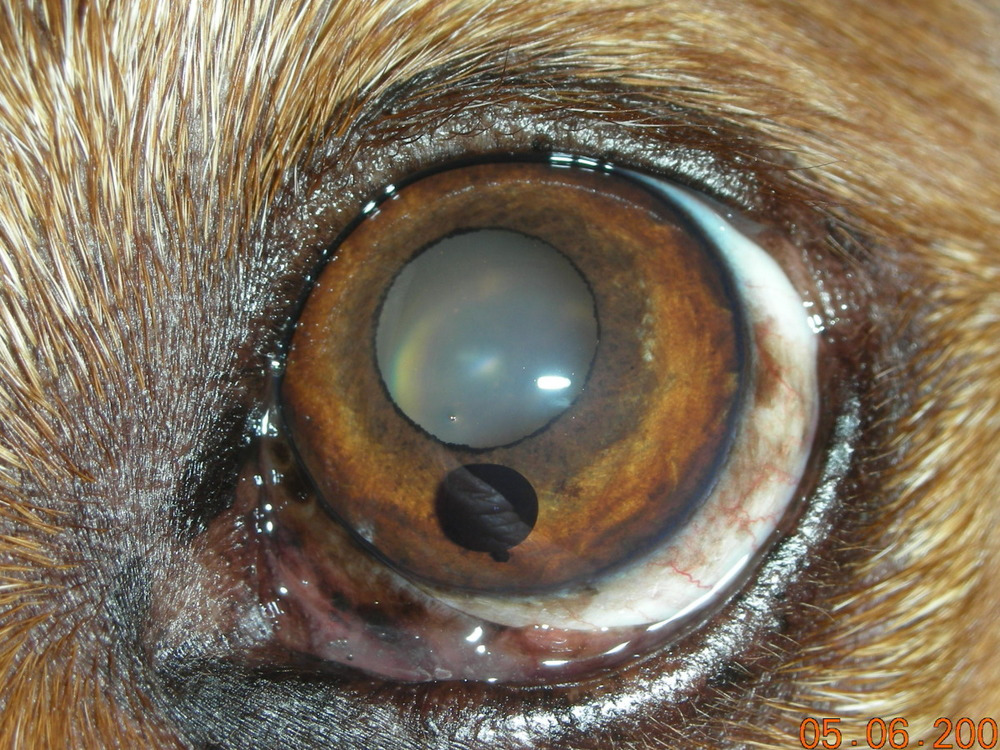
This is most commonly seen in the labrador retriever; These tumors appear as a densely pigmented, localized mass within the iris. The iris is most often affected, with the ciliary body and/or choroid affected secondarily by extension.
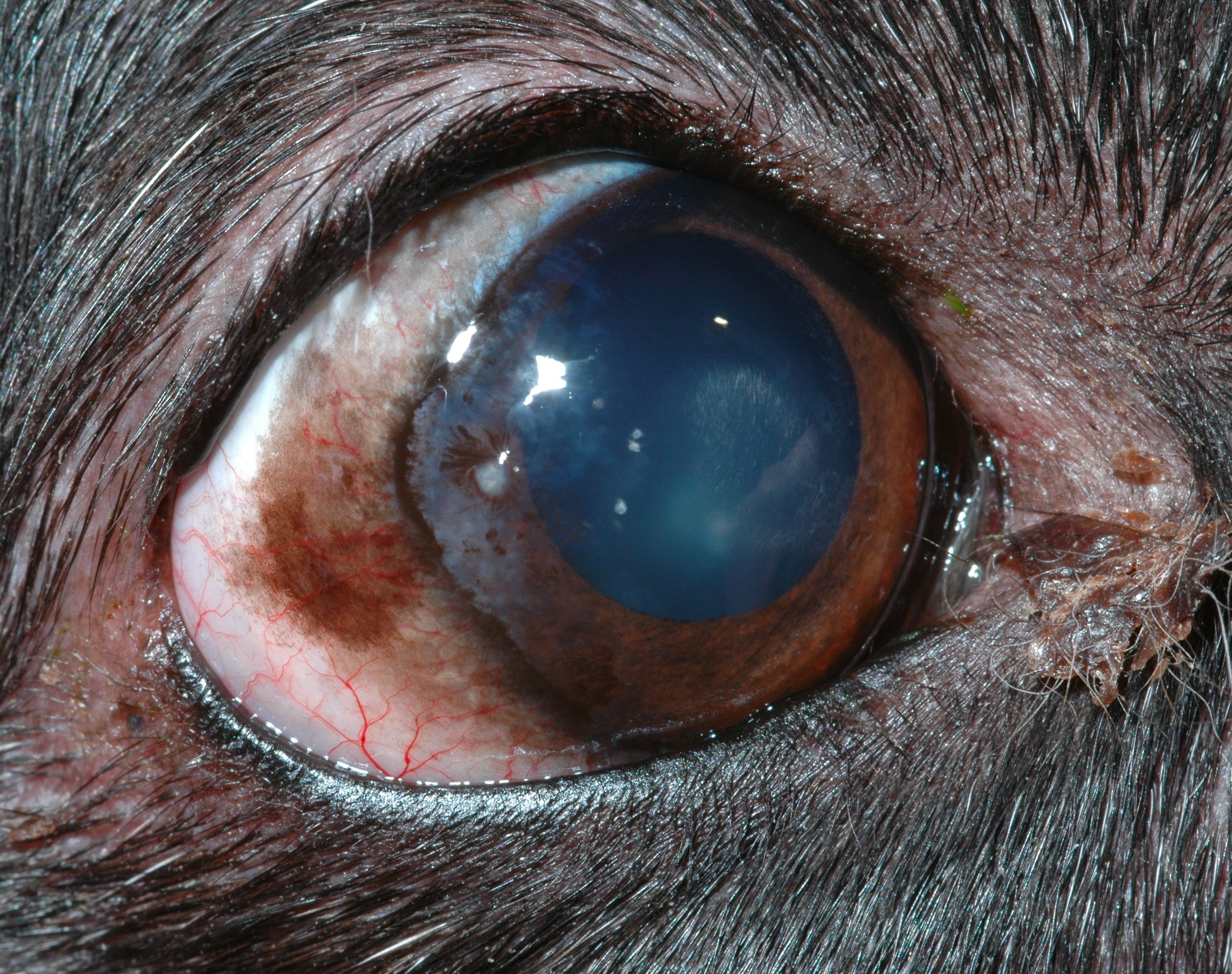
Primary choroidal melanoma is uncommon in dogs. On ophthalmic examination, hyphema, glaucoma, uveitis, iridal mass, and loss of vision were noted in the right eye. In dogs, uveal melanomas are generally benign with a very low incidence of metastasis.
Uveal melanomas represent the most common primary intraocular tumor. Within the ocular melanoma group, the uveal melanoma is characterized by being more aggressive than the epibulbar melanoma. Face (especially eyelids), trunk, and extremities;
Primary choroidal melanoma is extremely rare in dogs. Anterior uveal melanoma usually arise from anterior uveal tract (iris or ciliary body). Eye melanoma (ocular, uveal melanoma, melanoma of conjunctiva, eyelid melanoma) is a cancer that develops from melanocytes, cells that produce melanin pigment which is responsible for the color of the skin and eyes.
Clinically, dog eye melanoma is represented as an obvious mass in the eye along with hyphema (blood on the front surface of the dog eyes), glaucoma and pain. · in dogs, intraocular tumors typically arise from melanocytes in the root of or adjacent to the ciliary body (anterior uveal melanocytoma) · feline diffuse melanomas start as patchy iris hyperpigmentation that very slowly progresses to diffuse iris hyperpigmentation and thickening over several years; The eventual outcome is virtually always glaucoma
1 the good news is. The formation of a uveal, or iris, cyst is a common occurrence in dogs. Uveal melanomas are melanomas that may affect the dog's iris, ciliary body, and choroid.
Uveal melanocytic tumors may be benign (melanocytoma) or malignant (melanoma), with benign forms predominating in the dog. Intraocular melanomas are the most common primary intraocular tumors of dogs (2, 3). About 80% of uveal melanomas are benign (non
Recent studies shows that 4% of canine uveal melanomas metastasize or spread, and they do so within 3 months of being diagnosed with the condition. In cats, ocular melanoma initially presents as focal or multifocal areas of uveal pigmentation. These superficial areas confined to the surface of the iris are known as “iris freckles” or nevi.
These melanomas grow from the tissues that make up the uvea (the iris, ciliary body, and choroid). Melanoma is a very aggressive disease and growths are often large, regularly attacking the surrounding bones of the mouth prior to they are even identified by an owner or vet. However, skin tumors, particularly melanomas, are the most common malignant growths found in dogs.
In contrast, feline uveal cysts have been documented solely as benign with a breed predispostion for older burmese cats.

Vision Care For AnimalsGallery of Eye Diseases
Vision Care For AnimalsGallery of Eye Diseases
chronicsuperficialkeratitiscanine Animal Eye Clinic
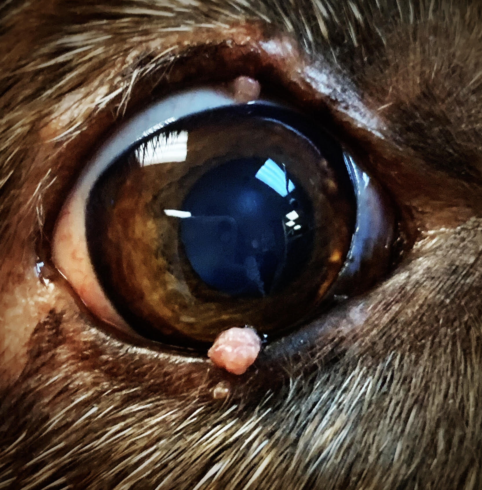
Eyelid Tumors The Sooner, The Better — ACVO Public
Uveal Melanomas Veterinary Ophthalmic Consulting